|
Imaging includes radiographs (x-rays) and sonograms (ultrasound images.) These diagnostic tools may be used by the veterinarian to assess the inner workings and structures of your cat. When used in conjunction with a physical examination and laboratory tests, imaging can give an in depth "picture" of your pet to help diagnose various diseases or conditions.
Cheshire Cat Feline Health Center is a full service hospital with high-quality imaging capabilities. Additionally we are able to partner with the Veterinary Imaging Center of San Diego to offer a full range of imaging services including consultation services, CT-Scan, MRI and more.
See their site at: Veterinary Imaging Center of San Diego

Radiography
Our state-of-the-art radiological equipment was recently replaced in order to provide higher quality images and more accurate results. Radiograpghs are produced when a low dose of x-rays (actually particles of light) are passed through the pet and onto a film cassette. The resulting image is really a picture of the shadows cast by the denser materials (like bones) in your cat's body. These shadows are projected onto a film that has been coated with a sensitive material. The film is developed in a manner very similar to a photograph.
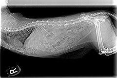
The image to the left shows a cat laying on it's side. The head (not captured in the image) is to the left and the tail is to the right. You can see the the bones, which are more dense, appear whiter in contrast to the less
dense muscles and organs. Radiographs are helpful in many cases such are when looking for a solid foriegn object that may have been swallowed, assessing broken bones or judging the size and shape of an organ. Because radiographs are only still images, ultrasound images may provide additional information when more answers are needed.
 Ultrasonography Ultrasonography
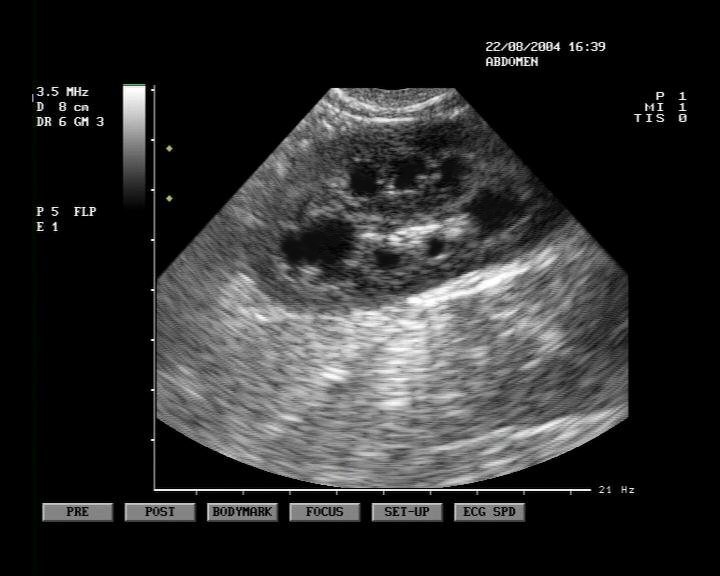
A sonogram, also known as an ultrasound, is a computerized picture taken by bouncing sound waves off organs and other interior body parts. A wand called a transducer is glided along the outside of the body over a centralized area or organ. As it glides, it introduces sound waves into the body. These sound waves bounce off the intended area and back into the transducer, which feeds the information into a computer. The picture then appears on a special computer screen.
|